The great feedback I received on this blog encouraged me to do more and expand the blog into a full website dedicated to data visualization in life sciences.
You can now find the blog at the following address:
www.gabrielaplucinska.com
On this new website you will find all of the previous blog posts, as well as data visualization I have made for #Makeover Monday, and resources on how to improve your science communication skills.
I hope that you will enjoy the new setup, and if you have any comments, criticism or suggestions let me know.
Figure Makeover
Monday, September 4, 2017
Tuesday, August 15, 2017
general organisation (aka use of white space)
So far in my posts I focused on single graphs and panels, but now I would like to tackle the general organization of an entire figure. Figures tend to be very crowded, a consequence of big amount of data and restrictions in number of figures. As a rule it is good to avoid busy figures, but it's rarely possible. Therefore, it's good to know some basic rules that improve readability and help keeping the flow of the story.
The key to effective storytelling through figures is the use of white space. It may sound silly, as we are mostly told to focus on the data itself, but white space is just as important.
Let's start with an example (Martorell-Riera et al. 2014)
the way this figure is organized makes you read it as follows:
After a moment you will realize that is not the order authors intended nor the one that makes sense. Another striking thing is general messiness (achieved by different scales and sizes of graphs/images) and a lot of white (non-used) space, which makes it look like panels were randomly put together.
Let't take it one panel at a time and then assemble them into a figure.
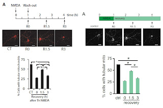
Panel A
I introduced color to the scheme to differentiate between different conditions. Additionally I took away the color from the images, so that even in this small version details became more visible.
At the same time this allowed me to remove most of the text. Finally, I cleaned the "significance bars" by removing all the extra ink.
Small changes:
-rearrangement of the blots vs graphs
-increase the size of blots, so they are easier to read.
You can now appreciate the addition of the color in the graph which corresponds to the condition from panel A's scheme.
Panel C
Similar changes as in the panel B. Also blue is now assigned to represent results of mitofusin2 knock down.
Added white space between the blots make the panel "breath", so it feels less crowded in small space.
 Panel D
Again: changes in images' look up table (LUT) and color added to the graph. Small changes in arrangement make better use of the available space and allow for bigger images.

Panel E
Adjusted LUT, added green bar to indicate which images represent neurons treated with NMDA (following color scheme from panel A).
A small detail that often escapes writers attention is "orientation" of the samples. In panel E all neurons are shown with their cell bodies at the bottom, but the Mfn2. This makes Mfn2 stand out from the rest, driving attention of the reader away from the actual phenotype. Small change - big impact.
 Panel F
I followed the same design rules and with small changes help the reader navigate through the graphs and relate them to certain conditions introduced at the beginning.
So now that we have all the elements, let's put them all together:
It becomes more obvious now that use of color helps the reader navigate through the figure and relate results to the treatment. Also the overall flow of the figure is more consistent, as all of the original data is followed by a quantification. Finally using white space more efficiently reduced the size of the figure without compromising its readability.
The key to effective storytelling through figures is the use of white space. It may sound silly, as we are mostly told to focus on the data itself, but white space is just as important.
Let's start with an example (Martorell-Riera et al. 2014)
the way this figure is organized makes you read it as follows:
After a moment you will realize that is not the order authors intended nor the one that makes sense. Another striking thing is general messiness (achieved by different scales and sizes of graphs/images) and a lot of white (non-used) space, which makes it look like panels were randomly put together.
Let't take it one panel at a time and then assemble them into a figure.
Panel A
I introduced color to the scheme to differentiate between different conditions. Additionally I took away the color from the images, so that even in this small version details became more visible.
At the same time this allowed me to remove most of the text. Finally, I cleaned the "significance bars" by removing all the extra ink.
Small changes:
-rearrangement of the blots vs graphs
-increase the size of blots, so they are easier to read.
You can now appreciate the addition of the color in the graph which corresponds to the condition from panel A's scheme.
Panel C
Similar changes as in the panel B. Also blue is now assigned to represent results of mitofusin2 knock down.
Added white space between the blots make the panel "breath", so it feels less crowded in small space.
 Panel D
Again: changes in images' look up table (LUT) and color added to the graph. Small changes in arrangement make better use of the available space and allow for bigger images.

Panel E
Adjusted LUT, added green bar to indicate which images represent neurons treated with NMDA (following color scheme from panel A).
A small detail that often escapes writers attention is "orientation" of the samples. In panel E all neurons are shown with their cell bodies at the bottom, but the Mfn2. This makes Mfn2 stand out from the rest, driving attention of the reader away from the actual phenotype. Small change - big impact.
 Panel F
I followed the same design rules and with small changes help the reader navigate through the graphs and relate them to certain conditions introduced at the beginning.
So now that we have all the elements, let's put them all together:
It becomes more obvious now that use of color helps the reader navigate through the figure and relate results to the treatment. Also the overall flow of the figure is more consistent, as all of the original data is followed by a quantification. Finally using white space more efficiently reduced the size of the figure without compromising its readability.
Friday, July 28, 2017
schematics [2]
It's summer and as usually things slow down, which is also reflected by my activity in writing new blog posts. In order not to be completely forgotten I prepared this short post about rather simple improvement on schematics. I have to say I really like making schemes as they give the most opportunities to show creativity.
This particular post is going to deal with schematics that overuse text (I actually wonder if they can still be called schematics then). I will show you how to nicely turn them into an effective graphic that is easy to comprehend at a first glance.
As an example I am going to use this scheme from Watanabe et al. 2016, where the only graphic authors used are arrows and boxes:
Such representation does the job and you can follow the protocol, but the text is rather small making it harder to read and you can easily call it boring, which will most likely cause reader to ignore it. Overall, it can be done more efficiently, with addition of some graphics:
1) I introduced centrifugation tubes making it easier to follow which part of the sample was used for further centrifugation.
2) Clearer organization and use of different font sizes (not too many though!) drives attention to more important elements
3) Removing all the unnecessary text gave the overall schematic more clarity and some "breathing" space.
I would like to finish this blog post with offering my help with making your schematics - do not hesitate to contact me with any questions you may have. I am happy to offer my advice and help out with your work.
Tuesday, June 20, 2017
on the move
One of the greater challenges in of showing data is illustration of the observed movement of particles, cells etc. These days you can submit videos alongside your manuscript, however some journals will ask you to refrain from it (e.g. Journal of Neuroscience) and your audience may not always be able to access it (or may consider it too much hustle). So until videos become the integral part of the pdf file, let's look at the options of depicting it in still images.
1) kymograph
A golden standard in the neuroscience field. Briefly, kymograph is a graphical representation of distance as a function of time. To researchers not familiar with it I like to show this graphic visualizing train transport, from 1885, which helps grasping a concept behind the kymograph.

Reason why this is a very powerful visualizing techniques is because you can read many transport parameters from it, like: direction of movement, speed of the particle, number and length of pauses, as illustrated in Marinković et al. 2012:

The main problem with the kymograph is that it works best when transport happens along one axis. If your particles are moving at multiple angles kymograph may not be the best solution.
2) single frames
This is the very classical approach to showing changes over time. Typically you split the movie into single frames and present them in vertical or horizontal order.
In those examples moving particles have been pseudocolored to focus attention on them. Alternatively you can use arrows to drive readers attention. This particular approach is commonly used when working with tracing, for example in cell migration.
3) overlay
This method is a variation of single frame representation and is very powerful when dealing with densely labeled samples. Basically, you overlay consecutive frames of the movie (at a selected interval) and pseudo-color them, in the following fashion:
frame 1 - blue
frame 2 - green
frame 3 - red
When you overlay those 3 frames, you will get the following image (Plucinska et al. 2012)
Stationary particles appear as white and the arrows point out to moving particles which appear in color (respectively to the coloring scheme above). If the time frames you choose are small and the environment is not too crowded you can see movement of individual particles. In this particular approach it is important to use primary colors (red, green, blue) - in the overlay they will appear as white, which is not the case if you use off colors.
4) cartoons
In paper by Marinković et al. authors had to deal with another problem. While they look at the axon in which transport happens bidirectianly, the density of the labeling would blur the overall images over-representing stationary mitochondria.
Therefore they chose to draw a line in the middle of the image (grey dotted line) and show how many mitochondria crossed it over time (additionally indicating the direction by green or magenta color).
To summarize, when you want to show your time lapse data, you have to consider following factors:
- movement directionality (bi-, multidirectional)
- labeling density
- speed of movement (generally slow moving particles will appear better as single frames, as you can put a "timer" on the images)
1) kymograph
A golden standard in the neuroscience field. Briefly, kymograph is a graphical representation of distance as a function of time. To researchers not familiar with it I like to show this graphic visualizing train transport, from 1885, which helps grasping a concept behind the kymograph.

Reason why this is a very powerful visualizing techniques is because you can read many transport parameters from it, like: direction of movement, speed of the particle, number and length of pauses, as illustrated in Marinković et al. 2012:
The main problem with the kymograph is that it works best when transport happens along one axis. If your particles are moving at multiple angles kymograph may not be the best solution.
2) single frames
This is the very classical approach to showing changes over time. Typically you split the movie into single frames and present them in vertical or horizontal order.
 |
| Misgeld et al.2007 |
In those examples moving particles have been pseudocolored to focus attention on them. Alternatively you can use arrows to drive readers attention. This particular approach is commonly used when working with tracing, for example in cell migration.
3) overlay
This method is a variation of single frame representation and is very powerful when dealing with densely labeled samples. Basically, you overlay consecutive frames of the movie (at a selected interval) and pseudo-color them, in the following fashion:
frame 1 - blue
frame 2 - green
frame 3 - red
When you overlay those 3 frames, you will get the following image (Plucinska et al. 2012)
Stationary particles appear as white and the arrows point out to moving particles which appear in color (respectively to the coloring scheme above). If the time frames you choose are small and the environment is not too crowded you can see movement of individual particles. In this particular approach it is important to use primary colors (red, green, blue) - in the overlay they will appear as white, which is not the case if you use off colors.
4) cartoons
In paper by Marinković et al. authors had to deal with another problem. While they look at the axon in which transport happens bidirectianly, the density of the labeling would blur the overall images over-representing stationary mitochondria.
Therefore they chose to draw a line in the middle of the image (grey dotted line) and show how many mitochondria crossed it over time (additionally indicating the direction by green or magenta color).
To summarize, when you want to show your time lapse data, you have to consider following factors:
- movement directionality (bi-, multidirectional)
- labeling density
- speed of movement (generally slow moving particles will appear better as single frames, as you can put a "timer" on the images)
Subscribe to:
Posts (Atom)


